
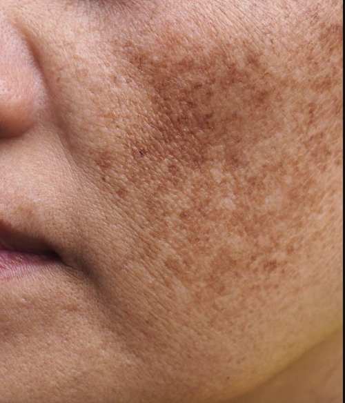
Melasma Causes and Treatment
Last Updated: August 8, 2026
Melasma is a chronic, acquired hypermelanosis characterized by symmetrical brown, grey-brown, or blue-grey macules and patches predominantly involving sun-exposed areas of the face, such as the forehead, cheeks, upper lip, and jawline[1]. Colloquially termed the “mask of pregnancy,” melasma represents one of the most clinically challenging pigmentary disorders encountered in dermatological practice worldwide[1].
While historically categorized strictly as an epidermal disorder of melanocyte hyperactivity, modern cutaneous biology demonstrates that melasma is a complex, photoaging-related pathology involving dermal vascularity, mast cell activation, basement membrane disruption, and systemic endocrine factors[2]. For patients seeking specialized care, understanding the full spectrum of causes, diagnostics, and medical treatments—such as those available through dermatological consultations in Kathmandu—is essential for long-term management.
💡 Clinical Takeaways
- A Whole-Skin Pathology: Melasma involves hyperactive melanocytes, increased dermal vascularization (VEGF expression), solar elastosis, and basement membrane damage[2].
- Beyond UV Rays: Triggers extend beyond ultraviolet radiation to include High-Energy Visible (HEV/blue) light, airborne particulate pollution, and neuroendocrine stress pathways[3, 4, 6].
- Targeted Diagnostics: In selected patients, comprehensive evaluation may require checking specific endocrine markers—including thyroid autoantibodies or reproductive hormones—to address underlying systemic drivers.
- Strict Limit on Combination Steroid Creams: Triple combination creams containing corticosteroids must be strictly limited to short-term, supervised use (typically 8–12 weeks maximum) to help prevent severe cutaneous atrophy and rebound pigmentation.
Contents
🚀 Advances in Melasma Research
Recent dermatological literature has expanded the treatment paradigm from simple pigment bleaching to comprehensive cellular repair:
- Opsin-3 (OPN3) Receptor Targeting: Research confirms that opsin-3 receptors on melanocytes sense visible blue light (400–500 nm), which may trigger prolonged pigment synthesis in darker skin types[3]. Tinted formulations containing iron oxides are widely recommended for visible light defense.
- Aryl Hydrocarbon Receptor (AhR) Activation: Airborne particulate matter (PM2.5) may activate AhR pathways in keratinocytes, inducing oxidative stress that upregulates tyrosinase activity[6]. Topical antioxidants are often integrated into standard regimens to combat pollution.
- Vascular-Melanocyte Paracrine Crosstalk: Endothelin-1 and VEGF secreted by dilated cutaneous capillary networks can sustain melanocyte hyperactivity[2]. Addressing underlying erythema via adjunctive treatments like Tranexamic Acid may help prevent relapse in selected patients.
Comprehensive Trigger Factors: The Scientific Mechanisms
Melasma melanocytes are hyper-reactive to internal and external micro-environmental changes. Identifying individual triggers allows for targeted lifestyle modification alongside medical therapy.
| Trigger Factor | Primary Source | Biological Pathway & Impact |
|---|---|---|
| Ultraviolet Radiation (UVA & UVB) | Sunlight, tanning units | Induces reactive oxygen species (ROS), mutates cellular DNA, and upregulates alpha-melanocyte-stimulating hormone (α-MSH), causing direct melanogenesis[1]. |
| Visible Blue Light (HEV) | Sunlight (primary), digital screens (secondary) | May stimulate Opsin-3 (OPN3) membrane receptors. While sunlight is the most significant source, prolonged screen exposure may contribute to oxidative stress in highly susceptible patients[3]. |
| Thermal Stress & Heat | Intense exercise, saunas, cooking heat | In experimental models, heat has been observed to activate temperature-sensitive channels (like TRPV3), which may promote pigment-related signaling[4]. |
| Psychological Stress & Sleep Loss | Chronic stress, circadian disruption | May influence the Hypothalamic-Pituitary-Adrenal (HPA) axis. Because ACTH shares a precursor with melanocyte-stimulating pathways, stress is considered a potential exacerbating factor[7]. |
| Air Pollution (PM2.5 / PAHs) | Traffic exhaust, industrial smoke | Polycyclic aromatic hydrocarbons may penetrate the stratum corneum, potentially activating Aryl Hydrocarbon Receptors (AhR) and contributing to inflammatory hyperpigmentation[6]. |
| Systemic Hormonal Shifts | Pregnancy, OCPs, HRT | Elevated estrogen (E2) and progesterone bind to specific cutaneous steroid receptors, increasing melanocyte transcription factors[1]. |
Medical Testing & Systemic Diagnostics
A precision dermatology approach involves objective clinical diagnostics. In specific cases—such as treatment-resistant melasma or when accompanied by other clinical symptoms—a dermatologist may consider targeted diagnostic testing to categorize pigmentation depth and rule out systemic exacerbators.
1. Cutaneous Imaging & Diagnostic Tools
- Wood’s Lamp Examination: Emits long-wave UVA (365 nm) light to accentuate epidermal pigmentation contrast. Epidermal melasma appears enhanced under Wood’s light, whereas dermal melasma shows less contrast.
- Dermoscopy: High-magnification surface microscopy evaluates pigment distribution (net-like vs. homogenous) and identifies underlying vascular patterns, such as telangiectatic vessels or diffuse background erythema[2].
- High-Frequency Ultrasound (HFUS): Non-invasive ultrasound imaging may be utilized in clinical settings to assess sub-epidermal low-echogenic bands (SLEB), helping to estimate epidermal-dermal thickness and solar elastosis.
2. Specific Endocrine & Blood Panels
When melasma presents atypically or fails to respond to standard care, a dermatologist may consider investigating systemic drivers in selected patients:
| Diagnostic Test | Specific Markers / Target | Clinical Rationale for Testing |
|---|---|---|
| Thyroid Function Panel | TSH, Free T3, Free T4, Anti-TPO Autoantibodies | Some clinical studies observe an association between autoimmune thyroid disorders and treatment-resistant melasma. Elevated TSH may enhance melanocyte responsiveness in certain patients. |
| Reproductive Hormone Panel | Serum Estradiol (E2), Progesterone, LH, FSH | Considered when evaluating luteal-phase hormonal surges or estrogen dominance that may influence steroid-driven pigmentation. |
| Serum Prolactin Test | Total Serum Prolactin | Hyperprolactinemia may stimulate melanocyte proliferation; typically checked in patients with galactorrhea or irregular menses. |
| Androgen Screening Panel | Total & Free Testosterone, DHEAS, SHBG | Screens for underlying conditions like Polycystic Ovary Syndrome (PCOS) when melasma is accompanied by adult acne, hirsutism, or alopecia. |
| Pelvic Ultrasonography (USG) | Ovarian morphology and cyst evaluation | Used in conjunction with androgen panels in selected patients to evaluate ovarian cysts and assist in ruling out or confirming PCOS. |
| Nutritional & Metabolic Panel | Vitamin B12, Vitamin D3 (25-OH), Serum Ferritin | Deficiencies in these markers are occasionally observed in chronic pigmentary disorders and may be assessed during a comprehensive workup. |
High-Altitude & Skin-of-Color Dynamics
Populations residing at higher elevations—such as the elevated topography of Nepal and mountain regions globally—face unique environmental physics. Ambient UV radiation generally increases by approximately 10% to 12% for every 1,000 meters of elevation gained. Combined with an increased prevalence of Fitzpatrick skin types III through V, high-altitude populations may experience accelerated melanogenesis and increased risk of post-inflammatory hyperpigmentation (PIH).
Additionally, the improper use of unregulated cosmetic bleaching agents or over-the-counter “fairness” formulations lacking medical oversight represents a major public health concern, often leading to barrier destruction and refractory dermal pigment deposition.
Evidence-Based Treatment Protocols
Successful management typically requires combining strict photoprotection with multi-targeted topical active ingredients, and in some cases, selective oral systemic agents or conservative clinical procedures.
1. Modern Photoprotection Standard
Photoprotection is universally recommended year-round. Dermatological guidelines suggest broad-spectrum SPF 50+ sunscreens providing high UVA protection. Formulations containing **iron oxides** (CI 77491, CI 77492, CI 77499) are often advised to help physically absorb and reflect visible light[3].
2. Topical Therapies
| Therapy | Mechanism of Action | Clinical Application & Safety Notes |
|---|---|---|
| Hydroquinone (2%–4%) | Competitively inhibits tyrosinase, decreasing conversion of DOPA to melanin[1]. | Gold-standard short-term therapy. Must be cycled under medical supervision to help prevent exogenous ochronosis or patchy hypopigmentation. |
| Triple Combination Cream | Combines Hydroquinone, Tretinoin, and a Topical Corticosteroid. | Highly effective for acute clearance. Strict short-term limitation applies (see warning below). |
| Azelaic Acid (15%–20%) | Selectively targets hyperactive melanocytes; exerts anti-inflammatory effects. | Often used as a long-term maintenance agent; generally considered safe during pregnancy and nursing. |
| Topical Tranexamic Acid (3%–5%) | Inhibits plasminogen activator, aiming to decrease arachidonic acid and prostaglandin levels. | May reduce both pigment synthesis and vascular erythema; often well-tolerated by sensitive skin. |
| Kojic Acid & Niacinamide | Kojic acid inhibits tyrosinase; Niacinamide may block melanosome transfer to keratinocytes. | Synergistic supportive ingredients commonly used in daily long-term maintenance formulations. |
⚠️ Critical Medical Warning: Short-Term Use of Triple Combination Creams
Triple combination creams (Hydroquinone + Retinoid + Topical Corticosteroid) are among the most effective prescription tools for acute melasma suppression[1]. However, the corticosteroid component makes long-term unmonitored use medically unsafe.
In many regions, patients unmonitored by dermatologists apply these creams continuously for months or years. Prolonged application of topical steroids on facial skin can lead to severe, localized complications:
- Topical Steroid-Damaged Face (TSDF) & Steroid Rosacea: Persistent facial redness, pustules, and severe burning sensations.
- Cutaneous Atrophy: Thinning of the dermis, rendering the skin fragile, translucent, and easily bruised.
- Telangiectasia: Permanently dilated, visible red blood vessels across the central face.
- Rebound Hyperpigmentation & Ochronosis: Severe, dark, blue-black discoloration that is far more resistant to treatment than original melasma.
Clinical Guideline: Prescription combination creams should generally be used for a maximum duration of 8 to 12 weeks under direct dermatological monitoring, followed by a transition to non-steroidal maintenance agents (such as Azelaic Acid).
3. Oral Systemic Therapies
In selected patients, off-label low-dose **Oral Tranexamic Acid (TXA)** may be considered as an adjunct to downregulate angiogenesis and melanogenesis[5]. Oral TXA requires careful pre-screening and is generally contraindicated in patients with personal or family histories of thromboembolic disease, deep vein thrombosis (DVT), significant renal impairment, or concomitant use of combined oral contraceptives[5].
4. In-Office Procedural Options
Procedural modalities may be considered when topical therapy alone is insufficient, selected carefully to avoid post-inflammatory hyperpigmentation (PIH):
- Superficial Chemical Peels: Low-concentration Mandelic Acid, Lactic Acid, or Glycolic Acid peels may gently accelerate epidermal turnover. Deep or aggressive peels are generally avoided due to inflammation risk.
- Low-Fluence Q-Switched Nd:YAG Lasers (Laser Toning): Sub-thermolytic, low-fluence laser toning aims to break up melanosomes without generating significant thermal damage. High-energy ablative lasers are typically contraindicated.
- Microneedling with Depigmenting Solution: Mechanical micro-perforations may be utilized to enhance the transdermal delivery of sterile active serums into the superficial dermis.
Frequently Asked Questions
Can psychological stress directly trigger a melasma flare?
Chronic stress may influence the Hypothalamic-Pituitary-Adrenal (HPA) axis. Because ACTH shares a precursor with melanocyte-stimulating pathways, stress is considered a potential exacerbating factor in some individuals[7].
Why is long-term use of fairness creams dangerous?
Many unregulated over-the-counter “fairness” or bleaching creams contain hidden, potent corticosteroids. Extended, unmonitored use can cause irreversible skin thinning, permanently dilated blood vessels (telangiectasia), steroid rosacea, and severe rebound hyperpigmentation.
Does computer or smartphone screen light worsen melasma?
While sunlight is the primary source of visible blue light, some researchers suggest prolonged, close-proximity screen exposure might contribute to oxidative stress in highly susceptible patients. However, daylight remains the most clinically significant trigger. A tinted sunscreen helps protect against visible light from all sources[3].
Why do dermatologists order thyroid blood tests for melasma?
Some clinical studies observe an association between autoimmune thyroid disorders and treatment-resistant melasma. In selected patients, a dermatologist may consider a thyroid function panel to help rule out systemic factors.
Can heat from cooking over a stove trigger melasma?
In experimental models, heat has been observed to activate temperature-sensitive channels (like TRPV3), which may promote pigment-related signaling. For some patients who consistently notice flares after cooking or heavy exercise, heat avoidance may be a helpful adjunct[4].
Are chemical peels safe for brown or darker skin tones?
Superficial chemical peels utilizing larger-molecule acids (such as Mandelic Acid) can be safe for darker Fitzpatrick skin types when selected carefully and performed by an experienced clinician. Pre-treatment skin priming and strict aftercare are typically required to help prevent post-inflammatory hyperpigmentation.
Is melasma permanently curable?
Melasma is medically categorized as a chronic, relapsing condition. While clinical treatments can achieve substantial fading of active lesions, maintenance therapy and strict daily photoprotection are necessary to help prevent relapse.
How long does clinical melasma treatment take to show results?
When responding to treatment, initial pigment lightening may be observed over 8 to 12 weeks of consistent therapy. However, timelines vary significantly depending on the individual, adherence to photoprotection, and the depth of pigmentation.
Schedule a Comprehensive Clinical Consultation
Managing chronic melasma requires accurate diagnostic classification, vascular assessment, and a strictly monitored medical plan. To receive an individualized treatment protocol tailored to your skin type, schedule a consultation at Derma Clinic.
Learn more about specialized melasma treatments in Kathmandu and book your clinical evaluation below.
Scientific References
- American Academy of Dermatology Association (AAD). Melasma Clinical Guidelines and Management Protocols.
- Masub N, Nguyen JK, Austin E, Jagdeo J. The Vascular Component of Melasma: A Systematic Review of Laboratory, Diagnostic, and Therapeutic Evidence. Dermatologic Surgery. 2020;46(12):1642–1650. doi:10.1097/DSS.0000000000002770
- Morgado-Carrasco D, et al. Melasma: The need for tailored photoprotection to improve clinical outcomes (Role of Iron Oxides and Visible Light). Photodermatology, Photoimmunology & Photomedicine. 2022;38(6):515–521. doi:10.1111/phpp.12783
- Zhang L, Zeng H, Jiang L, et al. Heat promotes melanogenesis by increasing the paracrine effects in keratinocytes via the TRPV3/Ca²⁺/Hh signaling pathway. iScience. 2023;26(5):106749. doi:10.1016/j.isci.2023.106749
- Godse K, et al. Oral Tranexamic Acid for the Treatment of Melasma: Evidence and Experience-Based Consensus Statement. Indian Journal of Dermatology. 2023;68(2):178–185. doi:10.4103/ijd.ijd_266_22
- Puri P, Nandar SK, Kathuria S, Ramesh V. Effects of air pollution on the skin: A review. Indian Journal of Dermatology, Venereology and Leprology. 2017;83(4):415-423. doi:10.4103/ijdvl.IJDVL_254_16
- Handel AC, Miot LD, Miot HA. Melasma: a clinical and epidemiological review. Anais Brasileiros de Dermatologia. 2014;89(5):771-782. doi:10.1590/abd1806-4841.20143063